A peer-reviewed publication by Drs. Raffaella Righetti and Md. Tauhidul Islam has reached the top 1% of cited publications in the field of clinical medicine over the last year. Righetti is an associate professor in the Department of Electrical and Computer Engineering at Texas A&M University. Islam, who was a doctoral student in the department at the time of publication, is now a physical science research assistant for the Stanford School of Medicine.
The paper, Noninvasive Imaging of Young’s Modulus and Poisson’s Ratio in Cancers in Vivo, was originally published in Scientific Reports, a publication of Nature. The research was conducted in the Ultrasound and Elasticity Imaging Laboratory, with the assistance of additional researchers from Texas A&M and Houston Methodist Hospital.
“We are very excited about our results so far,” Righetti said. “As the research is developed in the future, it could have major impacts on the diagnosis and treatment of many human diseases, including cancer.”
Righetti and Islam’s publication focuses primarily on developing imaging biomarkers that can be used by clinicians to determine a patient’s health. The researchers primarily focused on biomarkers of living tissue.
“In the last 30 years, there has been a great interest in developing noninvasive biomarkers,” Righetti said. “Biomarkers are measurements taken inside the body, usually with some type of imaging modality, that can tell us what’s happening in the body. When looking at tissues, we can tell if the tissue has a disease, how far the disease has progressed or if treatment methods are working.”
As the research is developed in the future, it could have major impacts on the diagnosis and treatment of many human diseases, including cancer.
For this study, Righetti and Islam were interested in the stiffness of tissue. Having an accurate and sensitive measurement of the stiffness of tissue is important for the medical field because it helps identify different diseases. The researchers focused primarily on cancer because the progression of cancer is known to change tissue stiffness.
“Cancers are typically harder than healthy tissue,” Righetti said. “This is why cancerous tumors are often discovered through palpation. However, the research we conducted is applicable to other diseases as well.”
For scientists to accurately measure the stiffness of tissue, they need two separate measurements, Young’s modulus (YM) and Poisson’s ratio (PR). YM is a mechanical parameter that is used as a measurement of the stiffness of tissue and PR is a measurement of the compressibility of the tissue. While it is difficult to measure these parameters noninvasively, Righetti and Islam’s method gives researchers an accurate measurement of both the YM and PR without an invasive procedure.
“This is the first study to noninvasively and simultaneously measure both the YM and PR,” Islam said. “Previous research only provided an estimate of the YM while assuming a predefined value of PR. This also made the estimate of the YM inaccurate because accurate assessment of YM requires knowledge of the actual PR.”
In addition to being the first study to accurately measure both YM and PR, Righetti and Islam’s method is also one of the first to treat tissue as a changing material.
“Most of the previous research assumed that tissues act as solids, but the behavior of cancers is more complex,” Righetti said. “Our model assumes that the tissues have more than one phase, solid and fluid, and essentially act like a sponge.”
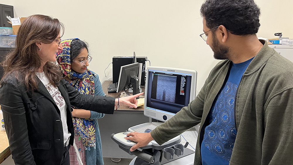
To measure the YM and PR, the researchers used an ultrasound imaging system to gather data while a small pressure was applied externally. They then processed the ultrasound data to determine internal mechanical changes in the tissue.
“Because we used an ultrasound, this method is very cost-effective,” Islam said. “However, the model we developed is broad enough to work with other imaging systems.”
To develop the model, Righetti and Islam began with Eshelby’s inclusion problem, a method used in soil sciences, and applied it to the measurement of tissue in the body. Using this method, they were able to apply a tested theory to modern biological science.
“We started with Eshelby’s inclusion problem because it is an established theory,” Islam said. “It has been tested in various areas of mathematics, engineering and biological sciences.”
While this research is the foundational step, the long-term ramifications could benefit many individuals around the world.
“This beginning step in our research is very promising,” Righetti said. “In the future, this method could be used to help determine the diagnosis and prognosis of diseases in the body. Once physicians begin treatment of a disease, they could also use it to monitor how the disease is changing in response to the treatment.”
Righetti hopes to expand the research to test the applicability of the model to the human body.
“Because it is cost-effective, this technology could provide an affordable tool to diagnose diseases in communities around the world,” Righetti said. “This opportunity to make a difference is what drives us to continue our research in the lab.”