“The goal of our work is to create a robust platform technology for label-free imaging of cellular and mitochondrial metabolism,” said Walsh. “This technology is useful for improving our knowledge of how metabolic heterogeneity within and across cell populations contribute to health, disease, and the discovery and evaluation of new drugs.
The potential benefits include a better understanding of how anesthesia drugs work and improved drugs and therapies for metabolic diseases such as cancer, diabetes and neurodegeneration.
Cellular-level metabolic information is necessary across multiple scientific disciplines, from nutrition and cancer biology to drug discovery. With this grant, Walsh hopes to advance autofluorescence technologies used for imaging cellular metabolism and robustly validate the technology and image analysis tools so that it can be easily adopted by other researchers.
She also hopes to use metabolic imaging technologies to evaluate the effects of anesthesia on cellular metabolism. There are currently gaps in scientific understanding of how anesthesia drugs function in cells and living organisms. A deeper understanding of these processes may lie within metabolic imaging.
The NIH MIRA R35 grant is awarded by the National Institute of General Medical Sciences to give investigators greater stability and flexibility by providing five years of funding for their research vision. The grant is awarded to a single principal investigator rather than several collaborators.
Rather than identifying specific research aims, the grant emphasizes the field that the individual researcher works in, the gaps present in that field and how the grant recipient is situated to address those gaps.
Walsh’s research areas include optical microscopy, label-free imaging, fluorescence lifetime and cellular metabolism.
Cellular metabolism
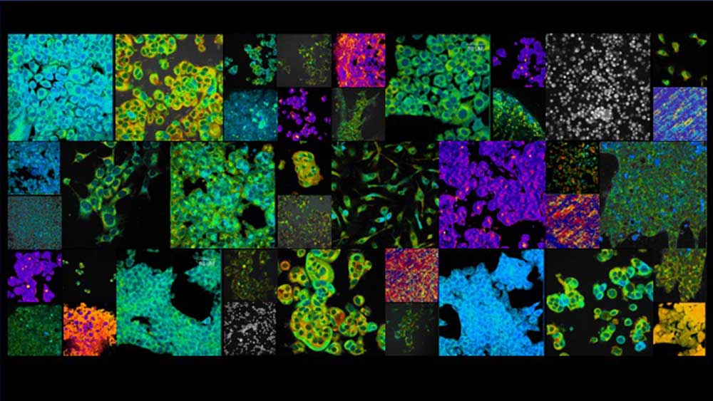
“Optical imaging means we use light to capture images of cells and tissues,” said Walsh. “Specifically, we look at cellular metabolism, which is the process that cells use to generate energy from sugar.”
During that process, cells use molecules called nicotinamide adenine dinucleotide (NADH) and flavin adenine dinucleotide (FAD). NADH and FAD serve as energy carriers so that reactions can occur. Both molecules, already present in cells, also have a physical phenomenon called fluorescence, where they give off light when shined with the light of the correct color wavelength.
Typically, fluorescence imaging requires fluorescent labels or dyes to provide contrast. Walsh uses the fluorescence of molecules innately within tissues, or autofluorescence, as contrast. She and her student researchers can detect differences in cells without adding contrast agents or dyes, as NADH and FAD provide the contrast.
This technology is further explained by a paper published in the journal JoVE. In addition to measuring the intensity or brightness of the fluorescence, Walsh and her team measure the fluorescence lifetime, or the time between the fluorescence photon absorption and emission.
Currently, autofluorescence lifetime imaging is excellent at detecting relative changes between two groups and can provide specific information about NADH and FAD enzyme-binding states, but pinpointing specific changes in metabolic pathways from the autofluorescence imaging is more complicated.
“The information we get in the imaging will change if cells use different substrates or different types of molecules to make energy,” she said. “If their rates change, then our quantitative information changes. There's still a gap for a robust model that can take what we measure with the label-free imaging and then predict what happened metabolically within the cell.
“The grant focuses on combining autofluorescence lifetime imaging with machine learning. We're trying to create models that can predict more specific metabolic information from the NADH and FAD fluorescence imaging metrics,” Walsh said.
Preliminary results
Since receiving the grant, Walsh and her doctoral student, Linghao Hu, have developed some exciting results with models that can predict whether a cell is using oxidative phosphorylation or glycolysis from the autofluorescence metrics.“It’s helpful if we can infer more information about metabolic pathway use or mitochondrial-specific metabolism from label-free imaging. We are working on computational models to achieve that goal. We are now testing the model across different cells and contexts and will soon try adding complexity to predict additional metabolic states,” said Walsh.
Since starting her research group in the biomedical engineering department at Texas A&M, Walsh has also received the Air Force Office of Scientific Research (AFOSR) Young Investigator Award and an AFOSR Defense University Research Instrumentation Program award to fund her research.
She is a Scialog Advancing BioImaging Fellow and this year, was awarded a collaborative Advanced Biomedical Imaging grant with Johannes Schöneberg from the Chan Zuckerberg Initiative Donor Advised Fund, an advised fund of the Silicon Valley Community Foundation.
She also serves as the Cain Development Faculty Fellow in the Department of Biomedical Engineering at Texas A&M.