Skip To Main Content
A collage of five art pieces featured in this article. In a maroon box are the words "Congratulations to the winners of the Artistic Engineering Contest."
The department's first annual Artistic Engineering art contest included submissions with images in multiple mediums. | Image: Texas A&M Engineering

The Department of Biomedical Engineering at Texas A&M University hosted its first annual Artistic Engineering art contest during the fall 2021 semester. Biomedical engineering-affiliated students, faculty and staff were invited to submit images in any medium and could include themes such as research, people in STEM, computer-generated imagery, photo composites or others.

Voting took place and the top five submissions were selected. These will be featured on the fifth floor of the Emerging Technologies Building.
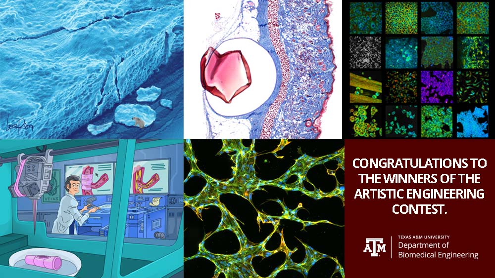
A microscope image of blue engineered living materials. The cells are stacked and look similar to an iceberg. In the corner of the image is a white polar bear.
Laura Rivera Tarazona designed this image using a scanning electron microscope to take photos of engineered living material from the lab. | Image: Courtesy of Laura Rivera Tarazona

First place: Laura Rivera Tarazona, doctoral student in Dr. Taylor Ware's lab, for

“Living Materials Save the Environment.” The glacier presented in this piece is an scanning electron microscope’s image of engineered living materials (ELM). The ELM material in this image consists of Escherichia coli contained within a hydrogel matrix. In the Ware Lab, they are synthesizing these ELMs to replace traditional plastics and help the global sustainability of natural environments.
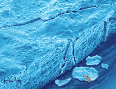
A cylindrical shaped cell under a microscope. On one side are small squares and rectangles dyed with blue and red ink. On the left is a large, heart shaped red piece of material.
This image was created by the Cardiovascular Pathology Laboratory, an interdisciplinary team of students and faculty in the College of Veterinary Medicine and Biomedical Sciences | Image: Courtesy of the Cardiovascular Pathology Laboratory
Second place: Members of the Cardiovascular Pathology Laboratory led by Dr. Fred Clubb for their work, “Serendipity: Trichrome,” a stained histology image of a subcutaneous polymer serendipitously shaped like a heart.
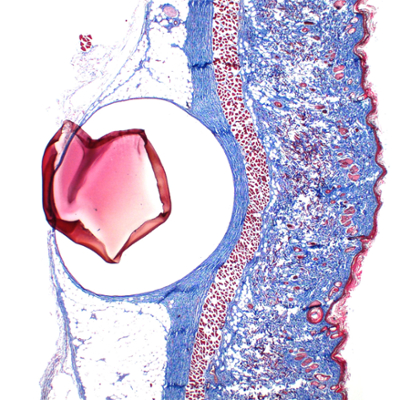
Sixteen boxes showing images of cells under a microscope. The clusters of cells are lit in a variety of colors ranging from blue and green to purple and pink.
Dr. Alex Walsh’s lab took these photos of fluorescence lifetime imaging to show the different active states of cells. | Image: Courtesy of the Quantitative Optical Imaging Lab

Third place: Members of the Quantitative Optical Imaging Lab led by Dr. Alex Walsh for

“Fluorescence Collage,” representative label-free fluorescence images of cell metabolism from the Quantitative Optical Imaging Lab. Fluorescence lifetime imaging is used to quantify the amount of time a fluorophore is in an excited state, which can enhance our understanding of certain microenvironmental changes. Contrast comes from molecules within cells, nicotinamide adenine dinucleotide (NADH) or flavin adenine dinucleotide (FAD), which are energy carriers used in metabolic reactions within the cell. Collage contains images of cancer cells, liver tissues, macrophages and organoids.
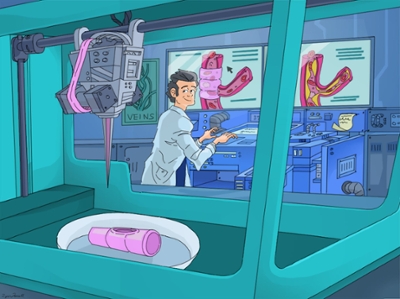
A cartoon showing a male scientist working in a lab. He is looking at a 3D printer that is printing a bio fabricated device. Behind the scientist is a screen showing that the device is meant to be placed around a blood vessel.
Ryan Farrell drew this cartoon to represent the bioprinting research taking place in several labs in the Department of Biomedical Engineering. | Image: Courtesy of Ryan Farrell
Fourth place: Ryan Farrell, master’s student in the Department of Visualization, who works with associate professor Dr. Akhilesh Gaharwar. “Bioprinting” is a cartoon that depicts the recent efforts of the Gaharwar and Dr. Abhishek Jain labs on 3D bioprinting blood vessels. These 3D bioprinted vessels mimic pathophysiological features of disease and health vascular tissue and can be used for drug testing.
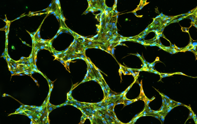
A series of thick and thin lines crossing a black background. The lines are dyed green and yellow with small blue circles throughout
Jim Tronolone’s research is focused on developing organs-on-chips, devices that allow researchers to create microenvironments outside of the body to test drug delivery and different disease treatments. | Image: Courtesy of Jim Tronolone
Fifth place: Jim Tronolone, doctoral student in Jain’s lab, for “Small Blood Vessels Forming Perfusable Networks in a Medical Device.” Blood vessels are a crucial component of most tissues of the human body, yet they are often neglected when engineers devise medical devices that model human tissues outside of the body. Such devices, called organs-on-chips, represent a new push towards advancing human in vitro research and replacing animal models for drug studies. This fluorescence micrograph demonstrates how an advanced organ-on-a-chip called a vascularized tissue-chip is fenestrated with small blood vessels called microvasculature. Such advanced devices are crucial for the translation of organ-chips to drug development and testing as well as basic biological investigation.