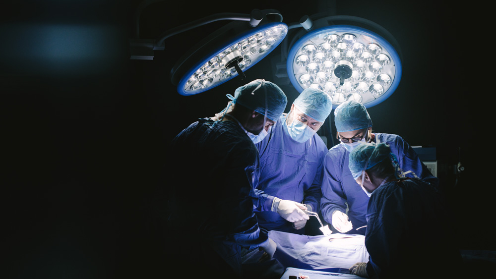
When someone breaks or fractures a bone, orthopedic surgeons can set and stabilize it by drilling and placing medical screws into the damaged area. This technique of fracture fixation enables fast healing and a quick recovery of functionality of the injured bone.
With extensive expertise needed, surgeons-in-training undergo residencies to hone their skills and bridge the gap between education and application under the mentorship of practicing doctors in a real-world setting.
Currently, this training consists of using either cadavers and simulant materials, such as wood or plastic, or virtual reality-based simulators to practice drilling and placing screws into a bone. However, as Dr. Bruce Tai explained, these methods do not imitate an actual, living patient very well nor do they provide realistic responses, and virtual reality systems are generally very expensive.
“Additionally, in both cases, the assessment and feedback given to trainees is totally subjective,” said Tai, assistant professor and Mechanical Engineering Industry Advisory Council Faculty Fellow in the J. Mike Walker ’66 Department of Mechanical Engineering at Texas A&M University. “I used to work with neurosurgeons to develop 3D-printed simulators and always felt that the assessment part was difficult (regardless of how good of a simulator they had), due to the lack of objective data.”
Merging engineering and medicine, a team of researchers were awarded the Simulation Research Grant from the Orthopaedic Research and Education Foundation (funded by the American Board of Orthopaedic Surgery). They are working to enhance the training of orthopedic residents through a 3D printed simulator and visualized performance data.
The team’s project aims to improve these training modules for future surgeons by combining the advantages of hands-on physical and objective, data-driven virtual simulations.
“This is a highly interdisciplinary project that brings together cognitive computing, spatial user interfaces and manufacturing to meet real-world challenges in medical training,” said Dr. Vinayak Krishnamurthy, mechanical engineering assistant professor. “To medical science, this project will lead to a holistic and objective approach to educate, train and evaluate medical students."
Physically, trainees will get firsthand experience drilling into and operating on 3D printed anatomical replicas made of a bone-mimicking composite previously developed by Tai that consists of a special printable plaster and binder jetting technology. This method enables different grades of hardness and toughness to be produced in a way that resembles human bones at different ages and conditions. Along with being inexpensive, 3D printing also allows students to print and interact with simulated rare diseases – giving them a hands-on experience with disorders that may not be present in available cadavers.
Virtually, data captured by embedded sensors will be used to analyze and assess the student’s performance as they conduct the simulated surgery. These sensors will record two key performance measures regarding drilling and screw placement: force and motion. As such, it will provide students and instructors alike an objective, real-time set of data from the simulator. That information can then be cross-referenced by a large dataset complied from experienced surgeons in order to objectively determine how well the operation was done.
“The outcomes of this project have the potential to lead to new intellectual insights,” said Krishnamurthy. “We could discover more about how humans process sensory information and apply it during high-pressure and risky tasks. Or gain insight about how to design new types of human-computer and human-machine interactions and interfaces to enable, expand and enhance human performance in such tasks. Finally, the project will also be able to shed light on fundamental techniques for manufacturing multiscale materials with a wide variety of physical properties to emulate biological objects.”
The team includes Tai, Krishnamurthy and Dr. Catherine Ambrose, associate professor at The University of Texas Health Science Center at Houston. The team is led by Dr. James Kellam, professor in the Department of Orthopedic Surgery at the UT Health Science Center at Houston.