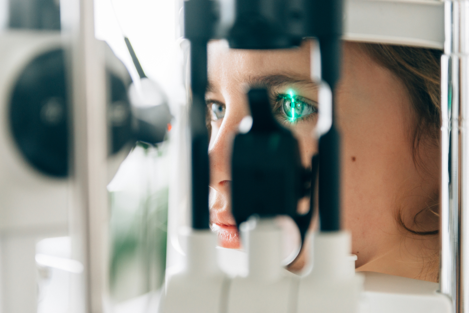
Doctors use a wide variety of imaging techniques to diagnose diseases. One such technique includes optical coherence tomography (OCT), which is a noninvasive medical imaging tool that has helped doctors accurately diagnose pathologies in cardiology, optometry and ophthalmology. The OCT technology was first introduced in 1991, and Texas A&M University researchers are set to give it a major upgrade.
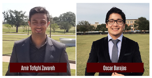
Graduate students Amir Tofighi Zavareh and Oscar Barajas, along with Dr. Sebastian Hoyos, associate professor in the Department of Electrical and Computer Engineering, have developed a new generation of OCT processing technology that will help doctors perform OCT tests faster and make a more accurate diagnosis. Low-complexity Asynchronous Re-sampler (LASR), their proprietary OCT processing technique, has an increased processing speed which helps doctors make clinical decisions based on high-resolution, real-time visualization.
“OCT is a noninvasive imaging technique capable of generating cross-sectional images of tissue architecture with high resolution,” said Tofighi Zavereh, the technical lead on the project. “The biggest challenge in current OCT technology was its slow scan speed, which gave an unsatisfactory image resolution. Doctors such as ophthalmologists need high-quality visualization for clinical decision making.”
The global OCT market is set to reach $1.6 billion in 2023 from just $140 million a year ago. Tofighi Zavareh and Barajas identified and addressed the needs of the fast-growing OCT market with their graduate research. The National Science Foundation’s I-Corp program awarded them a $50,000 grant to explore the commercial potential of their innovation with ophthalmology as a specific area of focus.
LASR will be implemented as a modular electronic device to integrate with an existing OCT instrument. The technology allows ophthalmologists to look at more eye tissues all in one scan.
“The LASR technology will help ophthalmologist better diagnose conditions of the eye, scan faster and potentially reduce the number of clinic visits,” said Barajas, the entrepreneurial lead. “All of these benefits will potentially help improve the vision of thousands of Americans in dire need of better diagnosis technologiesdirectly lower medical costs.”
The students, who are in the Analog and Mixed Signal Interfaces Laboratory in the electrical and computer engineering department, said their technology is based on a family of novel signal processing algorithms that significantly improve the accuracy and speed required for the medical images.
Tofighi Zavareh and Barajas’ I-Corps project, “Low-complexity Asynchronous Re-sampler for Optical Coherence Tomography,” has been three years in the making. They are collaborating with Dr. Xiaomin Yang, senior licensing manager in Texas A&M Office of Technology Commercialization and industry mentor, who helped them develop a business plan and get a provisional patent. They are also working with their co-industry mentor Dr. Saurabh Biswas, associate professor of practice in the Department of Biomedical Engineering and the Entrepreneur-in-Residence in TEES Division of Commercialization and Entrepreneurship and the McFerrin Center for New Ventures and Entrepreneurship in the Mays Business School to further develop their venture.
Learn more about their research here.